


Observation of Embryonic Fibroblasts with sCMOS Camera for Multichannel Imaging
SinceVision sCMOS cameras enable observation of embryonic fibroblasts through low light imaging and multichannel simultaneous imaging. Using high sensitivity, low readout noise, and up to 95% quantum efficiency, researchers can capture and analyze cytoplasm, mitochondria, and nuclear fluorescence signals with clarity.


Long-Term Observation of Embryonic Fibroblasts with Low-Light Imaging
Long-term observation of embryonic fibroblasts often means capturing migration, spreading, and division without damaging live cells. SinceVision sCMOS cameras solve the core conflict between image quality and cell health. With up to 95% light collection efficiency, these cameras capture weak fluorescence at lower excitation doses, reducing phototoxicity while improving quantitative data.
Researchers can run multichannel experiments over extended periods with stable, low-noise performance. This article shows how the SinceVision system produced clear, layered fluorescence images of cytoplasm, mitochondria, and nucleus in a single fibroblast sample, and explains why the technology is becoming a standard tool for live-cell imaging.
1. Experimental Background
Why Embryonic Fibroblasts Are Important Research Models
Embryonic fibroblasts are central to cell biology, developmental biology, and drug discovery. They are used extensively to study cell migration, spreading, cytoskeletal remodeling, and organelle function. Because these cells show clear morphological changes and strong adherent growth, they are ideal for dynamic structural observation and fluorescence imaging.
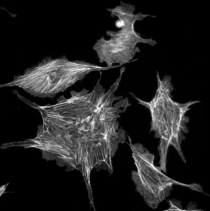
Figure: 1 Grayscale image of embryonic fibroblasts
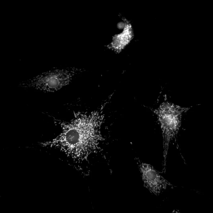
Figure 2: Grayscale image of embryonic fibroblast mitochondria
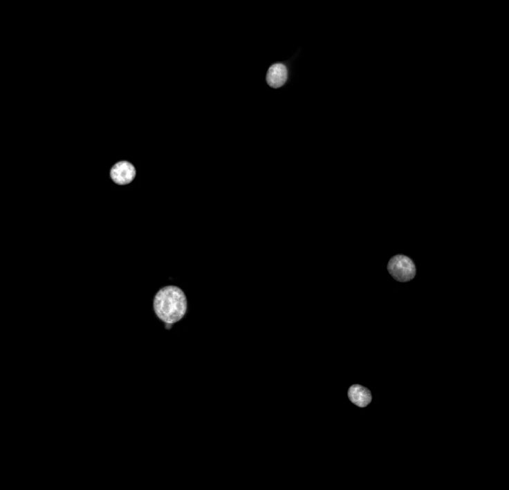
Figure 3: Grayscale image of embryonic fibroblast nuclei
The Challenge of Multichannel Imaging
Single-channel fluorescence staining reveals only one structure at a time. It cannot show functional relationships between different intracellular components. Researchers now need multichannel simultaneous imaging that delivers clear signal separation, distinct structural layers, and precise alignment across fluorescence channels. The key is a camera system that maintains sensitivity and low noise while switching between DAPI, FITC, and TRITC channels without damaging the sample.
2. Why sCMOS for Embryonic Fibroblast Imaging?
Traditional CCD cameras force a trade-off between speed and sensitivity. SinceVision sCMOS cameras use a back-illuminated sensor architecture that reaches 95% quantum efficiency at 560 nm. This means more photons become signal instead of noise, so excitation light can be kept very low. The result is less photobleaching and longer viable observation windows. The camera also holds a cooling temperature differential of at least 55 °C using a proprietary high-temperature soldering vacuum sealing process. Dark current and thermal noise are suppressed enough to make weak fluorescence signals usable across thousands of frames. For a researcher studying fibroblast organelle behavior over several hours, this stability is critical.
3. Imaging Setup and Results
How the Experiment Was Run
SinceVision's sCMOS camera was paired with a standard fluorescence microscope. Raw grayscale images were acquired for cytoplasm, mitochondria, and cell nucleus from the same field of view. The system captured fluorescence information from each cellular structure without saturating bright regions or losing dim signals. This laid a clean foundation for multichannel analysis.
Fluorescence Channel Processing
SinceVision's proprietary image processing software pseudo-colored the grayscale images to match the three standard fluorescence channels:
DAPI channel (blue) – nucleus
FITC channel (green) – cytoplasm
TRITC channel (red) – mitochondria
The pseudocoloring step preserved quantitative intensity relationships while making structural layers immediately identifiable.
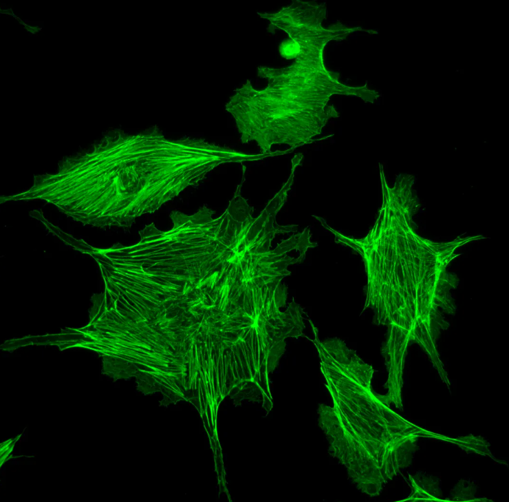
Figure 4: Pseudocolored image of embryonic fibroblasts green, FITC channel
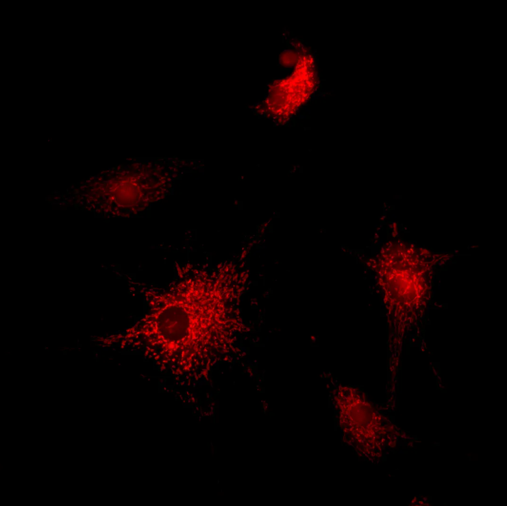
Figure 5: Pseudocolored image of embryonic fibroblast mitochondria (red, TRITC channel)
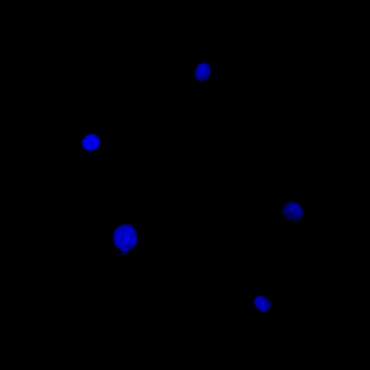
Figure 6: Pseudocolored image of embryonic fibroblast nuclei (blue, DAPI channel)
Multichannel Fusion Results
After processing, the three channels were overlaid into a single composite image. The fused image showed:
Blue signal (445 nm): precise nuclear localization
Green signal (530 nm): clear cytoskeletal filaments
Red signal (605 nm): accurate mitochondrial distribution
The composite offered clear structural layering, distinct separation between channels, and improved readability for analysis and publication. This same multichannel fusion approach can be applied to any combination of standard fluorophores that fall within the camera's high-QE window.
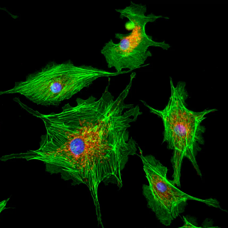
Figure 7: Fusion image of embryonic fibroblasts
4. SinceVision sCMOS Camera Advantages
Specification | Detail |
Sensor Type | Back-illuminated sCMOS |
Peak Quantum Efficiency | 95% at 560 nm |
Cooling Delta | ≥55 °C (proprietary vacuum sealing) |
Readout Noise | Low readout noise architecture |
Supported Channels | DAPI, FITC, TRITC (and other visible dyes) |
Software Integration | Multi-platform SDK, user-friendly UI |
High Sensitivity for Low Light Imaging
The back-illuminated sensor captures more photons per pixel. With 95% QE and a strong response across the visible spectrum, the camera reliably records weak fluorescence signals that other detectors might miss. This directly supports low light imaging applications where excitation light must be minimized.
Low Noise Design for Stable Multichannel Acquisition
Advanced thermal control and a low-readout-noise circuit suppress two major sources of image degradation. Even during long-term multichannel acquisition, dark current stays low and thermal noise remains controlled. Frame-to-frame consistency is high, which matters when tracking subtle organelle movements.
Easy Integration into Existing Workflows
The SinceVision sCMOS camera connects to standard microscopes without major modifications. A user-friendly software interface simplifies parameter setup, and the multi-platform SDK allows teams to embed image capture into existing analysis pipelines. Rapid deployment cuts the learning curve so researchers can focus on biology, not instrumentation.
5. Applications and Research Significance
The fibroblast imaging study demonstrates a clear research benefits:
High-definition structural evidence linking morphology and organelle function
Reliable visual assessment criteria for drug screening and toxicity testing
Support for long-term observation of cell migration and developmental mechanisms
Improved image quality for scientific presentations and publications
For any lab running live-cell fluorescence assays, the combination of high sensitivity, low noise, and multichannel support can improve throughput and data quality.
Contact SinceVision
SinceVision supports research laboratories worldwide with high-performance imaging tools. If you are working on low light imaging, multichannel fluorescence, or live-cell observation, reach out for product recommendations and a free sample trial.
Our team can help you configure the right sCMOS camera for your specific experimental needs and ensure that critical biological details stay clearly visible throughout your imaging sessions.
Frequently Asked Questions
What camera is best for live-cell low-light imaging of fibroblasts?
A back-illuminated sCMOS camera with high quantum efficiency and low readout noise. SinceVision's sCMOS reaches 95% QE and uses vacuum-sealed cooling to suppress dark current. This allows researchers to image weak fluorescence over hours without excessive photodamage.
How does the SinceVision sCMOS camera reduce phototoxicity during long-term observation?
High QE means fewer excitation photons are needed to generate a usable signal. The camera also maintains stable noise performance over time, so researchers can use lower light doses and still obtain clear images through the DAPI, FITC, and TRITC channels.
Can I use this camera for multichannel fluorescence of embryonic fibroblasts?
Yes. The camera's sensitivity covers the visible spectrum well, and the software supports pseudo-coloring and fusion of multiple channels. The fibroblast experiment successfully captured nucleus, cytoplasm, and mitochondria simultaneously with clear separation.
How can I get a sample trial of the SinceVision sCMOS camera?
SinceVision offers free sample trials for research labs. Contact the team through the website, describe your imaging challenge, and they will help you set up a trial with your own samples.
Featured Cameras
Solis B0465
Solis B518
You can also read
Observation of Embryonic Fibroblasts with sCMOS Camera for Multichannel Imaging
Jun 23, 2026
Automating Automotive Body Gap Detection with the SinceVision SR8060H 3D Laser Profiler
Jun 05, 2026
Curved Lens Inspection: How the Spectral Confocal Sensor Solves the Three Biggest Challenges
Jun 03, 2026
sCMOS Camera for Single-Photon Imaging | SinceVision's New Solis B0555 PRO
May 13, 2026
How Automotive Manufacturers Can Accurately Measure Conformal Coating Thickness on PCBA
Apr 29, 2026


 Compare
 Clear the comparison bar
