










OEM Integration of High-Speed sCMOS Cameras into Multimodal Fundus Imaging Devices
For multimodal fundus imaging devices, balancing near-infrared low-light sensitivity with the ultra-short exposure times required to prevent motion blur presents a critical engineering challenge. Integrating the SinceVision AIR Series high-speed camera and sCMOS camera provides a definitive solution.
 Product Inquiry
Fundus imaging devices must continuously capture high-resolution images of the fundus microvasculature, optic disc, and retina under near-infrared low-light conditions.
Due to microscopic eye tremors, involuntary blinking, and patient comfort limits, the effective image acquisition window is extremely short. The auxiliary light power of the system must also be strictly limited to protect the patient. Conventional industrial cameras fail in this complex operating environment, creating a technical dilemma: extending exposure time to capture weak light signals causes motion blur, while shortening exposure time to avoid motion blur results in severe image noise and a loss of diagnostic detail.
To capture physiological information accurately, the imaging system must achieve acquisition rates of thousands of frames per second while maintaining exceptional near-infrared sensitivity and stable data transmission.
Core Technical Requirements for Medical OEMs
Given the rigorous clinical applications of fundus imaging, the camera system integrated into the device must meet three primary technical benchmarks:
High-Speed Real-Time Acquisition: The target frame rate for raw image capture and transmission must reach 1,000 fps. High-density temporal sampling provides the exact data volume required for accurate multi-frame image fusion algorithms.
Near-Infrared Low-Light Sensitivity: The sensor must match the near-infrared auxiliary light operating band of the device. High near-infrared quantum efficiency is required to capture deep retinal tissue details without increasing the illumination intensity.
High Signal-to-Noise Ratio (SNR): Operating under ultra-short single-frame exposure conditions requires severe suppression of readout noise to guarantee image purity for intelligent diagnostic analysis.
System Configuration and Parameters
To meet the physical constraints of ocular imaging, the engineering configuration requires precise synchronization between illumination and capture.
Illumination System: The setup utilizes a narrow-band near-infrared LED light source with a wavelength of 820 to 850 nm, supporting pulsed or flickering operating modes. This penetrates retinal tissue while suppressing ambient stray light.
High-Frame-Rate Acquisition: The single-frame exposure time is restricted to a window of 0.5 to 1.0 milliseconds. Calibrating to a 1,000 fps capture rate guarantees that image blur caused by eye movements is mathematically minimized while shadow details in the histogram remain distinct.
Camera Selection: SinceVision AIR & Solis Series
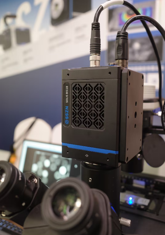
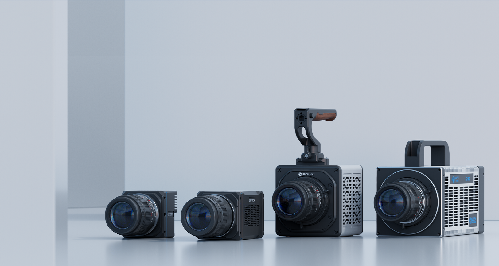
To solve the dual constraints of limited light and rapid motion, the recommended hardware integration is the SinceVision AIR Series high-speed camera and Solis Series sCMOS Camera.
The scientific cameras provides a compact engineering footprint that allows medical OEMs to customize the separation structure, thermal management systems, and SDK interfaces via C or E mount options.
Key Technical Specifications:
| Parameter | Specification | Engineering Benefit for Fundus Imaging |
| Max Resolution | 2560 × 2048 | Provides high-density raw data for multi-frame synthesis algorithms. |
| Frame Rate | 1,000 fps (Full Frame) | Eliminates motion blur from involuntary eye microtremors. |
| Pixel Size | 5.5 micrometers | Optimizes photon collection in dark optical environments. |
| Quantum Efficiency | 95% Peak (at 520 nm) | Back-illuminated sensor maximizes photon utilization in low-light conditions. |
| Readout Noise | 1.8 e⁻ | Balances diagnostic detail in both faintly reflected and high-brightness regions. |
| Dynamic Range | 89 dB | Prevents overexposure in the optic disc while retaining peripheral dark details. |
| Minimum Exposure | 1 microsecond | Allows for precise synchronization with pulsed LED illumination. |
Application Value and Clinical Results
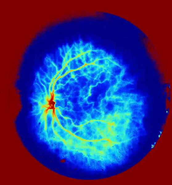
Integration testing demonstrates that the SinceVision AIR Series captures the full spectrum of ocular data without introducing saturated areas. Detail preservation remains exceptionally high even in bright regions like the optic disc, while the dark peripheral retinal areas retain full structural integrity.
The overall illumination distribution closely aligns with the stringent requirements of fundus optical imaging. By combining high speed, high sensitivity, and open integration capabilities, the AIR series & Solis series delivers stable, high-fidelity data input for multimodal fundus devices, providing a reliable foundation from initial photon acquisition to final algorithmic processing.






 Compare
 Clear the comparison bar
