










Multichannel Imaging of Embryonic Fibroblasts in Low Light
This application note highlights the use of SinceVision sCMOS cameras for the long-term, multichannel imaging of embryonic fibroblasts. By offering up to 95% quantum efficiency, the camera provides exceptional low light detection. This minimizes phototoxicity while capturing clear fluorescence signals from the cytoplasm, mitochondria, and nucleus, making it an essential tool for live-cell imaging.
 Product Inquiry
Related tags
Challenge of Live-Cell Imaging
Long-term observation of embryonic fibroblasts is essential in cell and developmental biology. However, capturing dynamic processes like cell migration and division requires observing these cells without damaging them.
Traditional imaging forces a difficult trade-off between image quality and cell health. Prolonged exposure to excitation light causes phototoxicity and photobleaching, while reducing the light often results in noisy, unusable data across multiple fluorescence channels.
Solution: High-Sensitivity sCMOS Technology
To solve the conflict between image quality and cell viability, researchers utilize the advanced sCMOS camera from SinceVision. Featuring a back-illuminated sensor architecture, this camera achieves up to 95% quantum efficiency at 560 nm.
This extreme sensitivity is critical for low light detection, allowing researchers to capture weak fluorescence signals using minimal excitation doses. By maximizing the conversion of photons into usable signals, the camera significantly extends viable observation windows for delicate live cells.
Multichannel Imaging Results
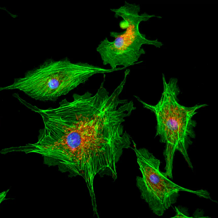
In this application, the SinceVision system was paired with a standard fluorescence microscope to observe embryonic fibroblasts. The camera successfully acquired clear grayscale images across three distinct channels:
DAPI channel (blue): Precise nuclear localization
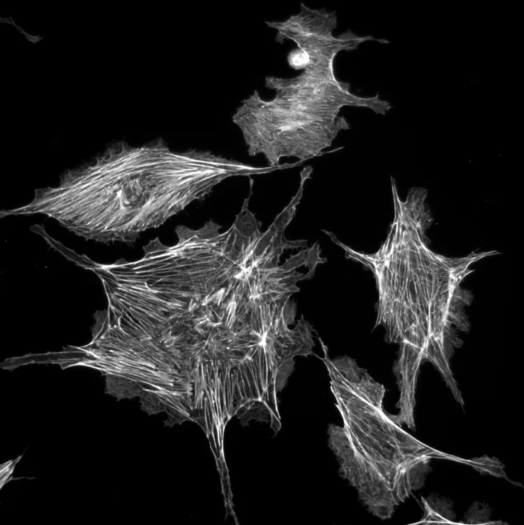
FITC channel (green): Clear cytoskeletal filaments in the cytoplasm
TRITC channel (red): Accurate mitochondrial distribution
Thanks to exceptional low light detection capabilities and a proprietary vacuum sealing process that maintains a cooling differential of at least 55 °C, the camera suppressed dark current and thermal noise. The resulting composite image delivered distinct structural layering and precise localization without saturating bright regions or losing dim signals.
Key Advantages for Research
Reduced Phototoxicity: Superior sensitivity ensures bright, quantifiable images at lower excitation doses, protecting cell health during extended studies.
Stable Multichannel Acquisition: Low readout noise and advanced thermal control guarantee frame-to-frame consistency during long, multi-fluorophore experiments.
Seamless Workflow Integration: A multi-platform SDK and user-friendly interface allow for rapid deployment into existing microscope setups.
Read the full experiment breakdown here






 Compare
 Clear the comparison bar
